
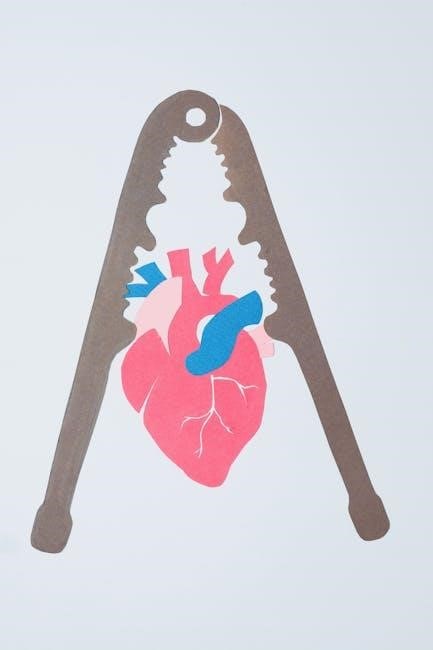
The heart is a vital organ located in the thoracic cavity‚ medially between the lungs. Its complex structure enables it to pump blood efficiently‚ sustaining life.
1.1 Overview of the Heart’s Structure
The heart is a muscular‚ hollow organ divided into four chambers: two atria and two ventricles. The septum separates the right and left sides. Blood flows through the heart via valves‚ ensuring one-way movement. The heart wall consists of three layers: the outer epicardium‚ the muscular myocardium‚ and the inner endocardium. These structures work together to pump blood efficiently through the circulatory system‚ maintaining life-sustaining functions.
1;2 Importance of Studying Heart Anatomy
Studying heart anatomy is crucial for understanding its role in circulation and overall health. It aids in diagnosing and treating cardiovascular diseases‚ which are leading causes of mortality. Knowledge of heart structure enables medical professionals to perform surgeries and develop treatments effectively. Understanding anatomy also helps in identifying congenital defects and managing conditions like hypertension. Additionally‚ it supports advancements in medical technology and personalized treatment plans. By studying heart anatomy‚ professionals gain insights into preventive measures and therapeutic interventions‚ ultimately improving patient outcomes and advancing cardiovascular medicine.

Gross Anatomy of the Heart
The heart is located in the thoracic cavity‚ medially between the lungs‚ within the mediastinum. Its structure includes chambers‚ valves‚ and surrounding tissues essential for function.
2.1 Location of the Heart in the Thoracic Cavity
The heart is centrally located in the thoracic cavity‚ within the mediastinum‚ a space between the lungs. It is encased in a double-layered membrane called the pericardium‚ which provides protection and support. The heart is positioned slightly to the left of the midline‚ with its base facing posteriorly toward the spine and its apex pointing anteriorly toward the chest wall. This strategic placement allows the heart to efficiently pump blood to the lungs and the rest of the body. The thoracic cage and surrounding organs‚ such as the trachea and esophagus‚ further stabilize its position‚ ensuring optimal functionality and protection from external forces.
2.2 Chambers of the Heart (Atria and Ventricles)
The heart consists of four chambers: two atria and two ventricles. The atria are the upper chambers that receive blood returning to the heart‚ while the ventricles are the lower chambers that pump blood out to the circulatory system. The right atrium receives deoxygenated blood from the body‚ and the right ventricle pumps it to the lungs. Conversely‚ the left atrium receives oxygenated blood from the lungs‚ and the left ventricle distributes it to the body. These chambers are separated by valves‚ ensuring blood flows in one direction. This arrangement allows the heart to efficiently manage the pulmonary and systemic circulations‚ maintaining proper oxygenation of the blood and supply to tissues throughout the body.
2.3 Heart Valves and Their Functions
The heart contains four valves that ensure blood flows in one direction through the chambers. The tricuspid valve is located between the right atrium and ventricle‚ while the pulmonary valve is between the right ventricle and pulmonary artery. Similarly‚ the mitral valve is situated between the left atrium and ventricle‚ and the aortic valve lies between the left ventricle and aorta. These valves open and close with each heartbeat‚ preventing backflow and ensuring efficient blood circulation. Proper valve function is critical for maintaining cardiac efficiency and overall health. Malfunctioning valves can lead to conditions such as stenosis or regurgitation‚ which may require medical intervention to restore normal heart function and prevent complications.
Microscopic Anatomy of the Heart
The heart’s microscopic anatomy reveals cardiac tissue types‚ including contractile cardiomyocytes and connective tissue. Cardiomyocytes contain myofibrils‚ enabling contraction‚ while intercalated discs ensure synchronized electrical activity across the heart muscle.
3.1 Cardiac Tissue Types
The heart is composed of three primary types of cardiac tissue: contractile‚ conductive‚ and connective. Contractile tissue‚ made of cardiomyocytes‚ is responsible for the heart’s muscular contractions. Conductive tissue‚ including the AV node and bundle of His‚ ensures the electrical impulses are transmitted efficiently‚ regulating heartbeat rhythm. Connective tissue‚ such as the endocardium and pericardium‚ provides structural support and protection. Each tissue type plays a crucial role in maintaining the heart’s functionality‚ ensuring proper blood circulation and overall cardiovascular health. Understanding these tissues is essential for diagnosing and treating heart conditions‚ as abnormalities in any tissue can lead to severe complications.
3.2 Cellular Structure of the Heart Muscle
The heart muscle‚ or myocardium‚ is primarily composed of cardiomyocytes‚ specialized striated muscle cells. These cells are uniquely designed for contraction‚ containing sarcomeres that enable rhythmic heartbeats. Cardiomyocytes are rich in mitochondria to meet high energy demands. Intercalated discs connect adjacent cells‚ facilitating synchronized contractions through gap junctions. Cardiac muscle fibers are organized into bundles‚ enabling efficient force generation. Additionally‚ the heart contains fibroblasts and endothelial cells‚ which support cardiac tissue by producing extracellular matrix and lining blood vessels‚ respectively. This complex cellular structure ensures the heart’s ability to pump blood continuously while maintaining structural integrity. Understanding this cellular anatomy is crucial for appreciating both normal heart function and disease mechanisms.

Blood Circulation Pathways
The heart manages two circulation pathways: pulmonary and systemic. Pulmonary circulation transports deoxygenated blood to the lungs‚ while systemic circulation delivers oxygenated blood to the body.
4.1 Pulmonary Circulation
Pulmonary circulation is a vital pathway where deoxygenated blood from the right ventricle flows through the pulmonary artery to the lungs. In the alveoli‚ oxygen diffuses into the blood‚ and carbon dioxide is removed. Oxygenated blood returns to the left atrium via the pulmonary veins‚ completing the cycle. This system operates under lower pressure compared to systemic circulation‚ ensuring efficient gas exchange. It is essential for maintaining oxygen supply and proper bodily functions. Understanding this pathway is crucial in diagnosing and treating conditions like pulmonary hypertension. Proper functioning of pulmonary circulation ensures the body receives the oxygen needed for energy production and overall health.
4.2 Systemic Circulation
Systemic circulation transports oxygen-rich blood from the left ventricle to the entire body and returns deoxygenated blood to the right atrium. The aorta‚ the largest artery‚ arises from the left ventricle and branches into smaller arteries‚ distributing blood to organs and tissues. Capillaries facilitate the exchange of oxygen‚ nutrients‚ and waste products. Deoxygenated blood collects in veins and returns to the heart through the superior and inferior vena cava. This system operates under higher pressure than pulmonary circulation‚ ensuring efficient delivery of oxygen to tissues. Proper functioning of systemic circulation is crucial for maintaining cellular function‚ energy production‚ and overall health. Any disruption can lead to conditions like hypertension or inadequate perfusion.

The Heart’s Electrical System
The heart’s electrical system regulates heartbeat rhythm. The sinoatrial node acts as the natural pacemaker‚ generating electrical impulses that control heart contractions and maintain circulation.
5.1 Cardiac Conduction System
The cardiac conduction system is a specialized network of cells that generates and transmits electrical impulses‚ regulating the heart’s rhythmic contractions. It begins with the sinoatrial (SA) node‚ the heart’s natural pacemaker located in the right atrium. From the SA node‚ impulses travel to the atrioventricular (AV) node‚ which delays the signal‚ allowing the atria to fully contract before the ventricles. The bundle of His carries the impulse further‚ dividing into the left and right bundle branches that stimulate the ventricles’ contraction. This system ensures coordinated and efficient pumping of blood‚ maintaining proper circulation throughout the body.
5.2 Role of the Autonomic Nervous System
The autonomic nervous system (ANS) plays a crucial role in regulating the heart’s activity. It consists of two branches: the sympathetic and parasympathetic systems. The sympathetic nervous system increases heart rate and contraction strength‚ preparing the body for physical activity. In contrast‚ the parasympathetic nervous system‚ primarily through the vagus nerve‚ promotes relaxation and reduces heart rate. This dual control allows the heart to adapt to changing conditions‚ ensuring efficient blood circulation. The ANS works in tandem with the cardiac conduction system‚ fine-tuning heart function based on the body’s needs. This intricate balance is essential for maintaining homeostasis and overall cardiovascular health.
Clinical Relevance of Heart Anatomy
Understanding heart anatomy aids in diagnosing conditions like coronary artery disease‚ valvular disorders‚ and cardiomyopathies. Accurate anatomical knowledge guides surgical interventions‚ improving patient outcomes and treatment success rates significantly.
6.1 Common Heart Conditions
The heart’s anatomy is crucial in understanding various clinical conditions. Coronary artery disease involves plaque buildup in arteries‚ while valvular disorders affect heart valve function. Cardiomyopathy weakens the heart muscle‚ disrupting blood flow. Arrhythmias‚ like atrial fibrillation‚ result from electrical system malfunctions. Heart failure occurs when the heart cannot pump blood adequately. Congenital defects‚ such as septal holes‚ are present from birth. These conditions highlight the importance of anatomical knowledge in diagnosis and treatment‚ enabling precise interventions to restore normal cardiac function and improve patient outcomes significantly.
6.2 Surgical Interventions
Understanding heart anatomy is essential for surgical interventions. Coronary artery bypass grafting (CABG) and heart transplants are common procedures for treating blocked arteries and end-stage heart failure. Valve repair or replacement surgeries address stenosis or regurgitation‚ often using bioprosthetic or mechanical valves. Minimally invasive techniques‚ like angioplasty with stent placement‚ are used for less severe blockages. Surgical interventions also include aneurysm repairs and congenital heart defect corrections. These procedures rely heavily on precise knowledge of the heart’s structure to ensure successful outcomes and restore normal cardiac function. Advances in surgical techniques continue to improve patient recovery rates and quality of life.
Educational Resources
Downloadable PDF guides‚ interactive tools‚ and platforms like TeachMeAnatomy offer detailed heart anatomy resources. Quizzes and 3D models enhance learning for students and professionals alike.
7.1 Recommended PDF Guides
Downloadable PDF guides provide comprehensive insights into heart anatomy. OpenStax’s Anatomy and Physiology offers detailed sections on cardiac structure. Kenhub’s heart anatomy PDF includes labeled diagrams and quizzes. 3D4Medical’s guides feature interactive 3D models for visual learning. TeachMeAnatomy provides PDFs with in-depth explanations and illustrations. These resources are ideal for medical students‚ educators‚ and professionals seeking to enhance their understanding. Additionally‚ platforms like Anatomy Lab and Study Unit offer downloadable worksheets and study guides. These PDFs are invaluable for self-study‚ offering both theoretical knowledge and practical exercises. They cater to diverse learning styles‚ ensuring a thorough grasp of heart anatomy.
7.2 Interactive Tools for Learning
Interactive tools enhance the learning of heart anatomy through engaging visual and kinesthetic experiences. Kenhub offers 3D heart models with zoom and rotation features‚ allowing detailed exploration of cardiac structures. TeachMeAnatomy provides quizzes and clickable diagrams to test knowledge and identify anatomical landmarks. 3D4Medical’s Complete Heart app features interactive simulations of heart functions‚ such as blood flow and valve operations. Virtual dissection tools enable users to explore layers of the heart digitally. These tools are particularly useful for medical students and educators‚ offering a hands-on approach to understanding complex anatomy. Quizzes and progress tracking further enhance learning outcomes‚ making them invaluable for self-study and classroom use.